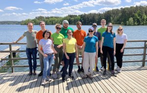
Ewolucja i genomika mikroorganizmów eukariotycznych
Tematyka badań
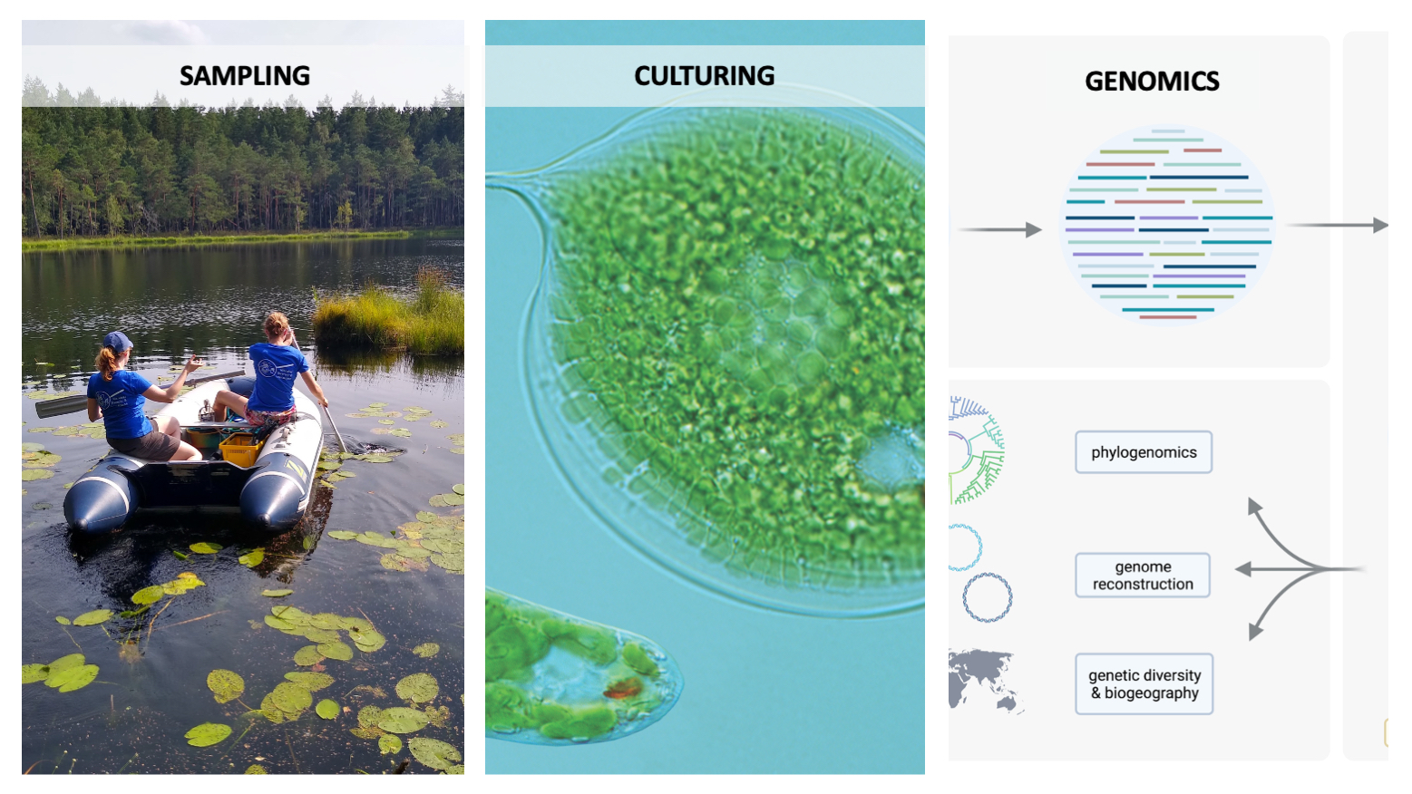
Prowadzone przez nas badania dotyczą różnorodności, ekologii i ewolucji mikroorganizmów eukariotycznych. Interesują nas zagadnienia ewolucji komórki eukariotycznej, w tym ewolucji organelli endosymbiotycznego pochodzenia, a także wpływu horyzontalnego transferu genów na ewolucję Eukaryota. Badania genomów i transkryptomów mikroorganizmów eukariotycznych pozwalają na poszukiwanie odpowiedzi na pytania dotyczące wczesnych etapów endosymbiozy, ewolucji genomów organellarnych oraz redukcji organelli. Staramy się również zrozumieć jaką rolę odgrywają mikroorganizmy eukariotyczne w środowiskach wodnych, poprzez badanie ich różnorodności i potencjału metabolicznego oraz ich interakcji z innymi mikroorganizmami.
W naszych badaniach wykorzystujemy zarówno techniki hodowli i izolacji komórek, jak również metody biologii molekularnej. Przede wszystkim jednak korzystamy z możliwości jakie niesie sekwencjonowanie nowej generacji i analizy bioinformatyczne. Pracujemy z danymi z sekwencjonowania genomów, transkryptomów, metagenomów czy też amplikonów. Rozwijamy również własne narzędzia bioinformatyczne przystosowane do pracy z danymi pochodzącymi z eukariota.
Projekty
Pochodzenie i ewolucja endosymbiozy: poszukiwanie stadiów przejściowych wśród mikroorganizmów eukariotycznych (SYMBIOSTART), Sonata BIS, Narodowe Centrum Nauki (kierownik: Anna Karnkowska)
Analiza różnorodności funkcjonalnej i taksonomicznej symbiontów wśród protistów z grupy Euglenozoa, Polonez BIS, Narodowe Centrum Nauki, (kierownik: Daria Tashyreva)
Fotosymbioza u słodkowodnych orzęsków: badanie różnorodności, funkcjonowania i ewolucji z zastosowaniem sekwencjonowania pojedynczych komórek, Grant Preludium BIS, Narodowe Centrum Nauki (kierownik: Anna Karnkowska)
Struktura i ewolucja genomów mitochondrialnych euglenin, Grant Preludium, Narodowe Centrum Nauki (kierownik: Paweł Hałakuc)
Zespoły mikroorganizmów słodkowodnych w gradiencie eutrofizacji: różnorodność i interakcje protistów i bakterii, Grant Opus, Narodowe Centrum Nauki (kierownik: Anna Karnkowska)
Evolution of phototrophy in eukaryotes, EMBO Installation Grant (kierownik: Anna Karnkowska)
Zakończone projekty
Ewolucja i funkcja odwróconych powtórzeń (IR) w genomach plastydowych Euglenophyta, Grant Preludium, Narodowe Centrum Nauki
Ewolucja i funkcja plastydów bezbarwnych glonów z grupy Euglenophyta i Dictyochophyceae, Grant Sonata
Współpraca
- Hampl lab, Uniwersytet Karola w Pradze, Czechy
- Solana lab, Exeter University, Anglia
- Kashiyama Lab, Fukui University of Technology, Japonia
- Kolisko lab, Institute of Parasitology, Ceske Budejovice, Czechy
- Fabrice Not, Sorbonne University, Francja
- Multicellgenome lab, Institute of Evolutionary Biology, Barcelona, Hiszpania
- Dr hab. Tomasz Jagielski, Uniwersytet Warszawski
- Dr Kacper Maciszewski, Biology Centre CAS, Czechy
Aktualności
19 marca
Nowy projekt w naszym zespole! Ania Karnkowska otrzymała grant NCN Sonata BIS na utworzenie zespołu, który będzie badał ewolucję endosymbiozy u mikroorganizmów eukariotycznych – SYMBIOSTART.
20 grudnia
Dr Ivan Garcia Cunchillos otrzymał prestiżowe stypendium EMBO postdoctoral fellowship! Gatulujemy i cieszymy się, że to właśnie z nami będzie próbował szukać odpowiedzi na pytania związane z wczesnymi etapami endosymbiozy plastydów.

1 listopada
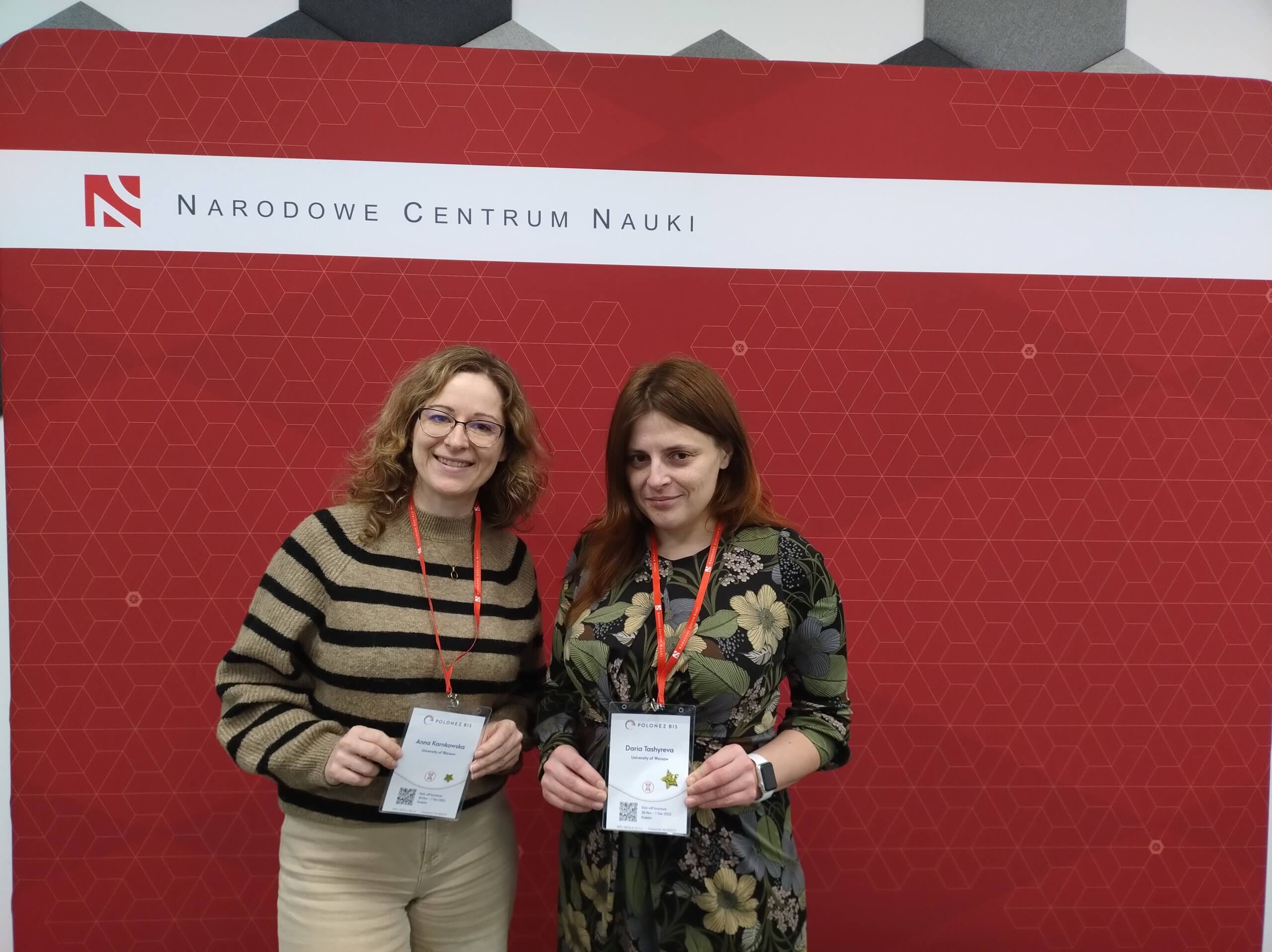
Witamy w zespole Dr Darię Tashyrevę, która będzie z nami pracować nad endosymbiontami Euglenozoa w ramach Jej grantu Polonez BIS 3 o akronimie SYMBIOZOANS!
5-7 października
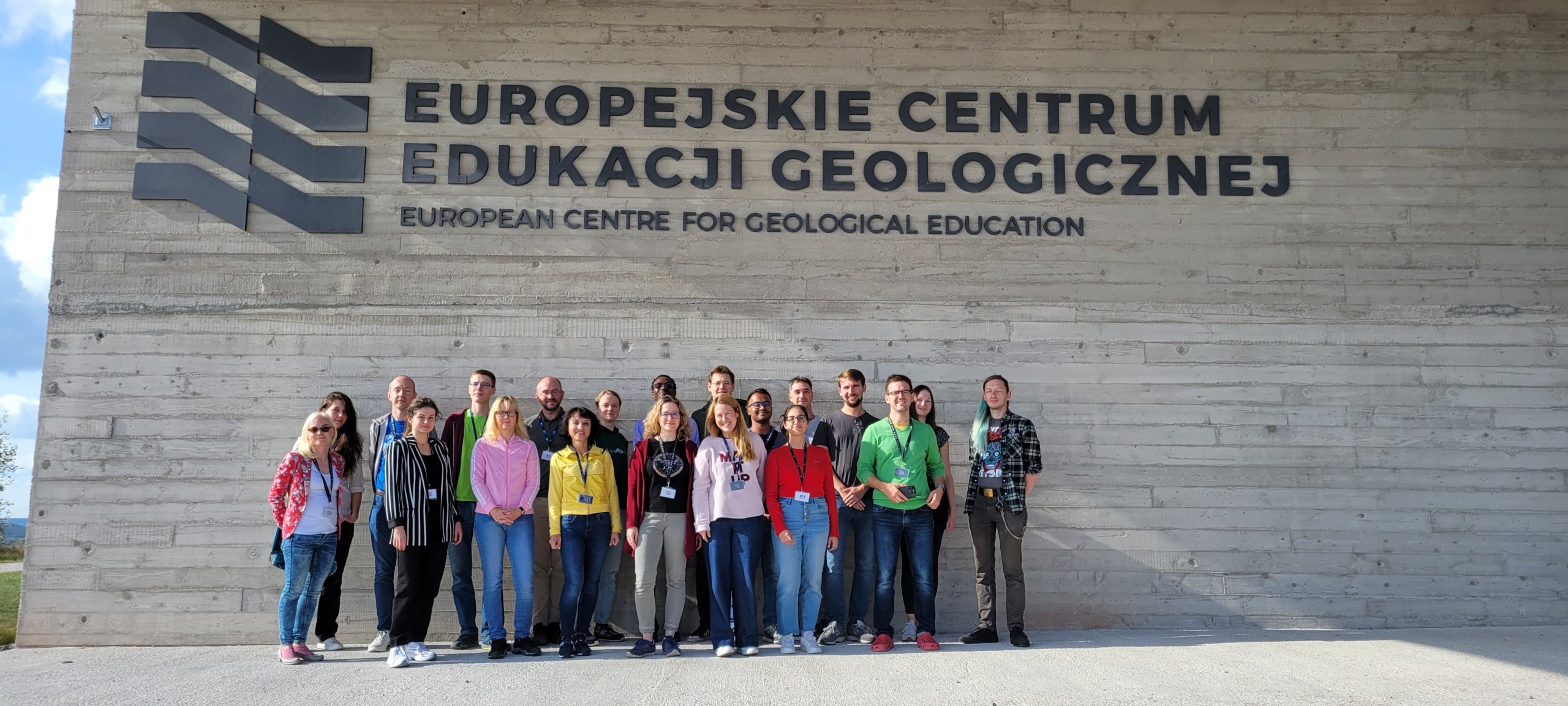
Bardzo udany wyjazd do Chęcin, na którym dyskutowaliśmy nasze projekty i plany z grupą Rafała Mostowego i Kasi Piwosz oraz zaproszonymi goścmi z Polski, Litwy i Chorwacji. Więcej o spotkaniu na stronie głównej IBE.
25 lipca – 6 sierpnia
Za nami kolejny udany wyjazd terenowy w ramach projektu MicroDivEr. Jak zwykle stacjonowaliśmy w Stacji WB w Urwitałcie.
1 lipca 2023
Witamy w naszym zespole Dr Anhelinę Kyrychenko, która w ramach prjketu EMBO Solidarity Grant będzie pracować z nami nad różnorodnością wirusów mikroorganizmów eukariotycznych.
3 czerwca 2023
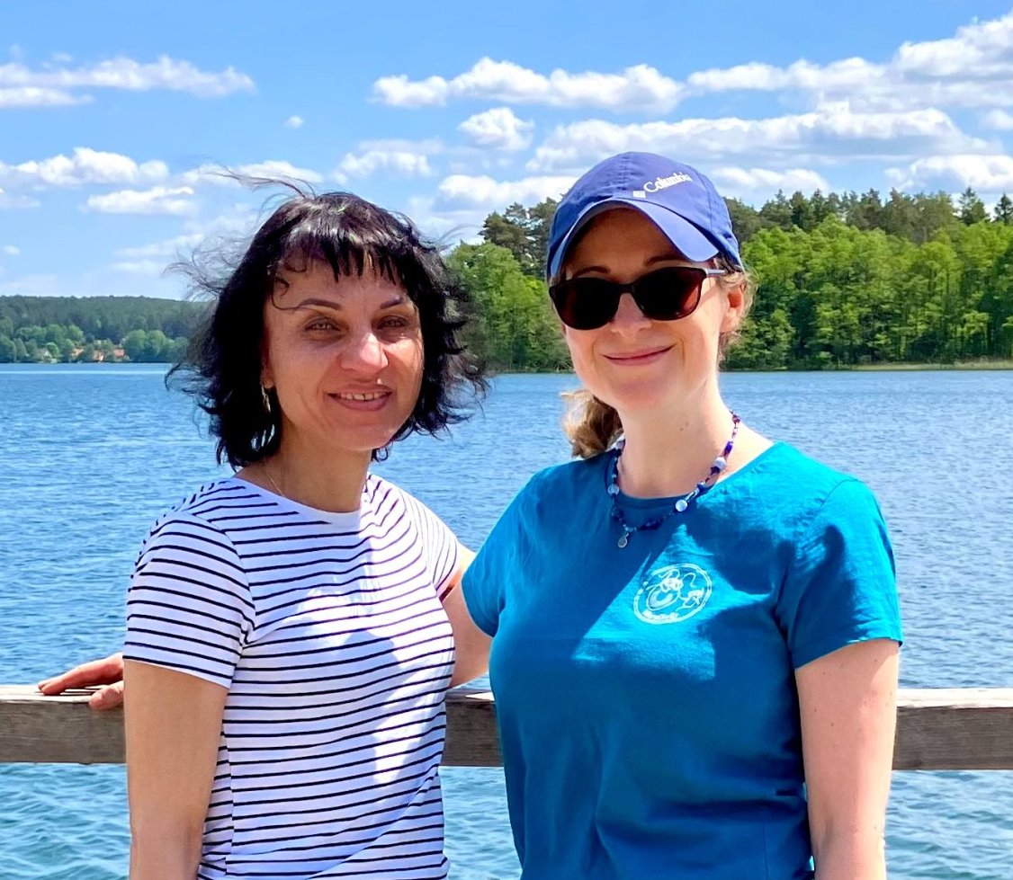
Ania odebrała dzisiaj oficjalnie swój dyplom habilitacyjny, a w zeszłym tygodniu awansowała również na stanowisko profesora uczelni!

31 maja – 2 czerwca
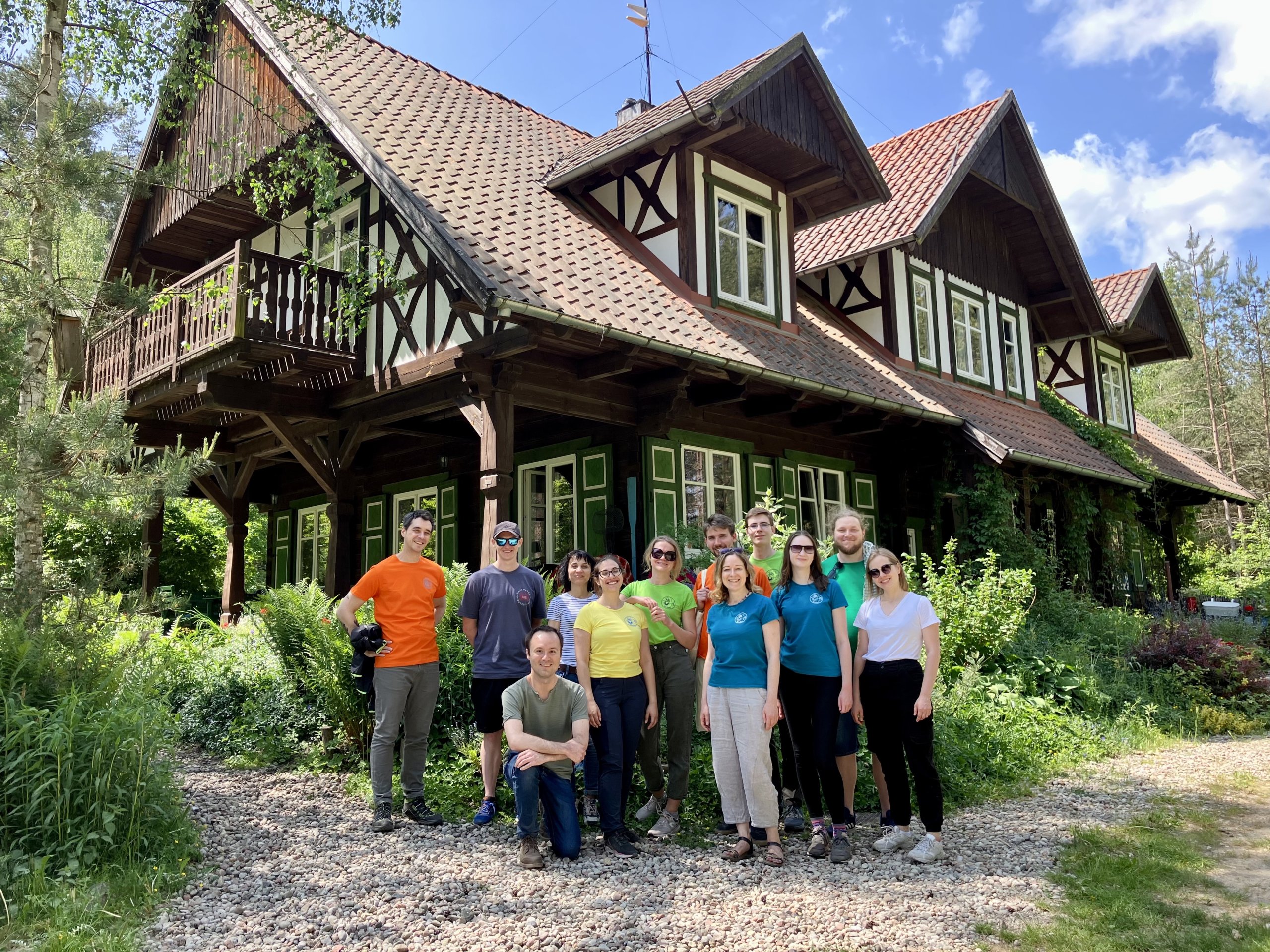
Za nami niezywkle udany lab retreat podczas którego nie tylko integrowaliśmy się z nowymi członkami zespołu, ale planowaliśmy też kolejne badania i dyskutowaliśmy co zrobić, żeby pracowało nam się jeszcze lepiej.
4 maja 2023
Do naszego zespołu, w ramach realizacji projektu MicroDivEr dołączył nowy post-doc dr Ivan Garcia Cunchillos. Witamy i cieszymy się na współpracę!
20 maja 2023
Nasza doktorantka, Valentina Smacchia, bierze udział w kursie Microbial metagenomics: a 360º approach w Heidelbergu! Udział Valentiny w kursie sfinansowany został z programu IDUB IV.4.1. Kompleksowy program wsparcia dla doktorantów UW
8 listopada 2022
Kacper Maciszewski obronił doktorat! Gratulujemy i życzymy dalszych sukcesów w trakcie stażu podoktorskiego w Czechach.
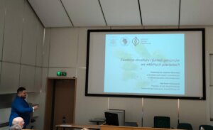
3 listopada 2022
Seda Mirzoyan,doktorantka na Uniwerytecie Karola w Pradze, odwiedziła nasz zespół w ramach realizacji wspólnego projektu finansowanego z mini grantu Sojuszu 4EU+ dla zespołów z udziałem UW.
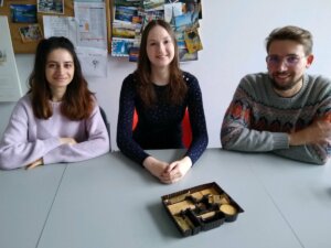
4 października 2022
Josiah Grzywacz, stypendysta Programu Fulbrighta dołączył do naszego zespołu na najbliższy rok akademicki, i będzie dalej z nami rozwijał swoje zainteresowania badawcze dotyczące fotosymbiozy u orzęsków.
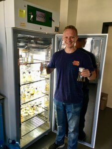
1 października 2022
Nowa doktorantka w naszym zespole! Mgr Małgorzata Chwalińska będzie realizować projekt w projekcie NCN Preludium BIS.
12 września 2022
Konkurs na stanowisko student-stypendysta w projekcie MicroDivEr. Termin nadsyłania zgłoszeń upływa 23 września. Więcej szczegółów w ogłoszeniu.
1 września 2022
Projekt pt. Opracowanie metody wysokoprzepustowego sekwencjonowania transkryptomów z pojedynczych komórek mikroorganizmów eukariotycznych uzyskał finansowanie ramach konkursu Nowe Idee, Inicjatywa Doskonałości Uczelnia Badawcza
1 maja 2022
W czasopiśmie Molecular Phylogenetics and Evolution właśnie ukazała się najnowsza publikacja naszego zespołu pt. Maturyoshka: A maturase inside a maturase, and other peculiarities of the novel chloroplast genomes of marine euglenophytes.
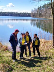
28 kwietnia 2022
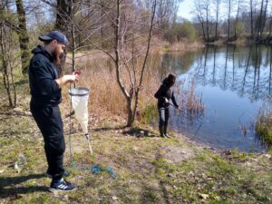
Za nami pierwszy w tym roku wyjazd terenowy na Mazury w ramach projektu #MicroDivEr. Jeziora jak zawsze piękne, a zespół w świetnych nastrojach!
19 kwietnia 2022
Konkurs na stanowisko doktorant-stypendysta w projekcie MicroDivEr. Termin nadsyłania zgłoszeń upływa 6 maja. Więcej szczegółow w ogłoszeniu.
13 kwietnia 2022
Witamy w zespole nowego pracownika technicznego Jakuba Kalinowskiego! Kuba już oswaja się z tajnikami pracy z mikroorganizmami eukariotycznymi i wziął udział w naszym pierwszym wiosennym wypadzie po próby.
9 kwietnia 2022
Z lekkim opóźnieniem świętujemy Dzień Darwina na Wydziale Biologii. W ramach tego wydarzenia Ania Karnkowska przybliży zagadnienia ewolucji komórki eukaiotycznej. Zapraszamy!
6 kwietnia 2022
Gratulujemy dr hab. Annie Karnkowskiej, której grant NCN Preludium BIS uzyskał finansowanie i będzie realizowany w naszym zespole.
30 marca 2022
Projekt Monitoring diversity of microbial dark matter in expanding anoxic zones uzyskał mini grant Sojuszu 4EU+ dla zespołów z udziałem UW. Projekt będziemy realizowali z zespołem z Uniwerystetu Karola i Sorboną.
28 marca 2022
Na kanale youtube Wydziału Biologii w ramach serii rozmów z kobiuetami nauki ukazał się wywiad z Anią Karnkowską.
1 marca 2022
Witamy Kacpra Maciszewskiego! Kacper wrócił z półrocznego pobytu na Uniwersytecie Alberty w grupie badawczej prof. Joela Dacksa realizowanego w ramach stypendium NAWA im. Iwanowskiej.
14 stycznia 2022
Już jest dostępna najnowsza publikacja naszego zespołu opisująca narzędzie Tiara, które służy do klasyfikacji sekwencji metagenomowych. Zachęcamy do korzystania!
21 grudnia 2021
Gratulujemy dr hab. Annie Karnkowskiej stypendium NAWA im. Bekkera, które umożliwi jej wyjazd na staż naukowy do IBE Barcelona!
26 listopada 2021
Gratulujemy mgr Pawłowi Hałakucowi, którego grant NCN Preludium uzyskał finansowanie i będzie realizowany w naszym zespole!
1 października 2021
Witamy w zespole nową doktorantkę Valentinę Smacchia!
Przykładowe publikacje
Maciszewski, Kacper; Wilga, Gabriela; Jagielski, Tomasz; Bakuła, Zofia; Gawor, Jan; Gromadka, Robert; Karnkowska, Anna
Reduced plastid genomes of colorless facultative pathogens Prototheca (Chlorophyta) are retained for membrane transport genes Journal Article
In: BMC Biology, vol. 22, no. 1, pp. 294, 2024, ISSN: 1741-7007.
@article{maciszewski_reduced_2024,

title = {Reduced plastid genomes of colorless facultative pathogens Prototheca (Chlorophyta) are retained for membrane transport genes},

author = {Kacper Maciszewski and Gabriela Wilga and Tomasz Jagielski and Zofia Bakuła and Jan Gawor and Robert Gromadka and Anna Karnkowska},

url = {https://doi.org/10.1186/s12915-024-02089-4},

doi = {10.1186/s12915-024-02089-4},

issn = {1741-7007},

year  = {2024},

date = {2024-12-01},

urldate = {2024-12-01},

journal = {BMC Biology},

volume = {22},

number = {1},

pages = {294},

abstract = {Plastids are usually involved in photosynthesis, but the secondary loss of this function is a widespread phenomenon in various lineages of algae and plants. In addition to the loss of genes associated with photosynthesis, the plastid genomes of colorless algae are frequently reduced further. To understand the pathways of reductive evolution associated with the loss of photosynthesis, it is necessary to study a number of closely related strains. Prototheca, a chlorophyte genus of facultative pathogens, provides an excellent opportunity to study this process with its well-sampled array of diverse colorless strains.},

keywords = {},

pubstate = {published},

tppubtype = {article}

}
Hollender, Metody; Sałek, Marta; Karlicki, Michał; Karnkowska, Anna
Single-cell genomics revealed Candidatus Grellia alia sp. nov. as an endosymbiont of Eutreptiella sp. (Euglenophyceae) Journal Article
In: Protist, vol. 175, no. 2, 2024, ISSN: 1434-4610.
@article{Hollender2024,

title = {Single-cell genomics revealed Candidatus Grellia alia sp. nov. as an endosymbiont of Eutreptiella sp. (Euglenophyceae)},

author = {Metody Hollender and Marta Sałek and Michał Karlicki and Anna Karnkowska},

doi = {10.1016/j.protis.2024.126018},

issn = {1434-4610},

year  = {2024},

date = {2024-04-01},

urldate = {2024-04-00},

journal = {Protist},

volume = {175},

number = {2},

publisher = {Elsevier BV},

abstract = {Though endosymbioses between protists and prokaryotes are widespread, certain host lineages have received disproportionate attention what may indicate either a predisposition to such interactions or limited studies on certain protist groups due to lack of cultures. The euglenids represent one such group in spite of microscopic observations showing intracellular bacteria in some strains. Here, we perform a comprehensive molecular analysis of a previously identified endosymbiont in the Eutreptiella sp. CCMP3347 using a single cell approach and bulk culture sequencing. The genome reconstruction of this endosymbiont allowed the description of a new endosymbiont Candidatus Grellia alia sp. nov. from the family Midichloriaceae. Comparative genomics revealed a remarkably complete conjugative type IV secretion system present in three copies on the plasmid sequences of the studied endosymbiont, a feature missing in the closely related Grellia incantans. This study addresses the challenge of limited host cultures with endosymbionts by showing that the genomes of endosymbionts reconstructed from single host cells have the completeness and contiguity that matches or exceeds those coming from bulk cultures. This paves the way for further studies of endosymbionts in euglenids and other protist groups. The research also provides the opportunity to study the diversity of endosymbionts in natural populations.},

keywords = {},

pubstate = {published},

tppubtype = {article}

}
Kolisko, Martin; Maciszewski, Kacper; Karnkowska, Anna
Plastid Evolution in Non-photosynthetic Lineages Book Chapter
In: Endosymbiotic Organelle Acquisition, pp. 203–237, Springer International Publishing, 2024, ISBN: 9783031574467.
@inbook{Kolisko2024b,

title = {Plastid Evolution in Non-photosynthetic Lineages},

author = {Martin Kolisko and Kacper Maciszewski and Anna Karnkowska},

doi = {10.1007/978-3-031-57446-7_7},

isbn = {9783031574467},

year  = {2024},

date = {2024-00-00},

urldate = {2024-00-00},

booktitle = {Endosymbiotic Organelle Acquisition},

pages = {203--237},

publisher = {Springer International Publishing},

keywords = {},

pubstate = {published},

tppubtype = {inbook}

}
Campo, Javier; Carlos-Oliveira, Maria; Čepička, Ivan; Hehenberger, Elisabeth; Horák, Aleš; Karnkowska, Anna; Kolisko, Martin; Lara, Enrique; Lukeš, Julius; Pánek, Tomáš; Piwosz, Kasia; Richter, Daniel J.; Škaloud, Pavel; Sutak, Robert; Tachezy, Jan; Hampl, Vladimír
The protist cultural renaissance Journal Article
In: Trends in Microbiology, 2023, ISSN: 0966-842X.
@article{DELCAMPO2023,

title = {The protist cultural renaissance},

author = {Javier Campo and Maria Carlos-Oliveira and Ivan Čepička and Elisabeth Hehenberger and Aleš Horák and Anna Karnkowska and Martin Kolisko and Enrique Lara and Julius Lukeš and Tomáš Pánek and Kasia Piwosz and Daniel J. Richter and Pavel Škaloud and Robert Sutak and Jan Tachezy and Vladimír Hampl},

url = {https://www.sciencedirect.com/science/article/pii/S0966842X23003281},

doi = {https://doi.org/10.1016/j.tim.2023.11.010},

issn = {0966-842X},

year  = {2023},

date = {2023-12-14},

urldate = {2023-01-01},

journal = {Trends in Microbiology},

abstract = {Protists are key players in the biosphere. Here, we provide a perspective on integrating protist culturing with omics approaches, imaging, and high-throughput single-cell manipulation strategies, concluding with actions required for a successful return of the golden age of protist culturing.},

keywords = {},

pubstate = {published},

tppubtype = {article}

}
Novák, Lukáš V. F.; Treitli, Sebastian C.; Pyrih, Jan; Hałakuc, Paweł; Pipaliya, Shweta V.; Vacek, Vojtěch; Brzoň, Ondřej; Soukal, Petr; Eme, Laura; Dacks, Joel B.; Karnkowska, Anna; Eliáš, Marek; Hampl, Vladimír
Genomics of Preaxostyla Flagellates Illuminates the Path Towards the Loss of Mitochondria Journal Article
In: PLOS Genetics, vol. 19, no. 12, pp. 1-30, 2023.
@article{10.1371/journal.pgen.1011050,

title = {Genomics of Preaxostyla Flagellates Illuminates the Path Towards the Loss of Mitochondria},

author = {Lukáš V. F. Novák and Sebastian C. Treitli and Jan Pyrih and Paweł Hałakuc and Shweta V. Pipaliya and Vojtěch Vacek and Ondřej Brzoň and Petr Soukal and Laura Eme and Joel B. Dacks and Anna Karnkowska and Marek Eliáš and Vladimír Hampl},

url = {https://doi.org/10.1371/journal.pgen.1011050},

doi = {10.1371/journal.pgen.1011050},

year  = {2023},

date = {2023-12-07},

urldate = {2023-12-07},

journal = {PLOS Genetics},

volume = {19},

number = {12},

pages = {1-30},

publisher = {Public Library of Science},

abstract = {The notion that mitochondria cannot be lost was shattered with the report of an oxymonad Monocercomonoides exilis, the first eukaryote arguably without any mitochondrion. Yet, questions remain about whether this extends beyond the single species and how this transition took place. The Oxymonadida is a group of gut endobionts taxonomically housed in the Preaxostyla which also contains free-living flagellates of the genera Trimastix and Paratrimastix. The latter two taxa harbour conspicuous mitochondrion-related organelles (MROs). Here we report high-quality genome and transcriptome assemblies of two Preaxostyla representatives, the free-living Paratrimastix pyriformis and the oxymonad Blattamonas nauphoetae. We performed thorough comparisons among all available genomic and transcriptomic data of Preaxostyla to further decipher the evolutionary changes towards amitochondriality, endobiosis, and unstacked Golgi. Our results provide insights into the metabolic and endomembrane evolution, but most strikingly the data confirm the complete loss of mitochondria for all three oxymonad species investigated (M. exilis, B. nauphoetae, and Streblomastix strix), suggesting the amitochondriate status is common to a large part if not the whole group of Oxymonadida. This observation moves this unique loss to 100 MYA when oxymonad lineage diversified.},

keywords = {},

pubstate = {published},

tppubtype = {article}

}
Hehenberger, Elisabeth; Boscaro, Vittorio; James, Erick R.; Hirakawa, Yoshihisa; Trznadel, Morelia; Mtawali, Mahara; Fiorito, Rebecca; Campo, Javier; Karnkowska, Anna; Kolisko, Martin; Irwin, Nicholas A. T.; Mathur, Varsha; Scheffrahn, Rudolf H.; Keeling, Patrick J.
In: Journal of Eukaryotic Microbiology, vol. 70, no. 5, pp. e12987, 2023.
@article{Hehenberger2023,

title = {New Parabasalia symbionts Snyderella spp. and Daimonympha gen. nov. from South American Rugitermes termites and the parallel evolution of a cell with a rotating “head”},

author = {Elisabeth Hehenberger and Vittorio Boscaro and Erick R. James and Yoshihisa Hirakawa and Morelia Trznadel and Mahara Mtawali and Rebecca Fiorito and Javier Campo and Anna Karnkowska and Martin Kolisko and Nicholas A. T. Irwin and Varsha Mathur and Rudolf H. Scheffrahn and Patrick J. Keeling},

url = {https://www.scopus.com/inward/record.uri?eid=2-s2.0-85162158230&doi=10.1111%2fjeu.12987&partnerID=40&md5=fa6e625d9458584ab89cd3c3f0943aa3},

doi = {10.1111/jeu.12987},

year  = {2023},

date = {2023-06-06},

urldate = {2023-01-01},

journal = {Journal of Eukaryotic Microbiology},

volume = {70},

number = {5},

pages = {e12987},

abstract = {Most Parabasalia are symbionts in the hindgut of “lower” (non-Termitidae) termites, where they widely vary in morphology and degree of morphological complexity. Large and complex cells in the class Cristamonadea evolved by replicating a fundamental unit, the karyomastigont, in various ways. We describe here four new species of Calonymphidae (Cristamonadea) from Rugitermes hosts, assigned to the genus Snyderella based on diagnostic features (including the karyomastigont pattern) and molecular phylogeny. We also report a new genus of Calonymphidae, Daimonympha, from Rugitermes laticollis. Daimonympha's morphology does not match that of any known Parabasalia, and its SSU rRNA gene sequence corroborates this distinction. Daimonympha does however share a puzzling feature with a few previously described, but distantly related, Cristamonadea: a rapid, smooth, and continuous rotation of the anterior end of the cell, including the many karyomastigont nuclei. The function of this rotatory movement, the cellular mechanisms enabling it, and the way the cell deals with the consequent cell membrane shear, are all unknown. “Rotating wheel” structures are famously rare in biology, with prokaryotic flagella being the main exception; these mysterious spinning cells found only among Parabasalia are another, far less understood, example.},

keywords = {},

pubstate = {published},

tppubtype = {article}

}
Žárský, Vojtečh; Karnkowska, Anna; Boscaro, Vittorio; Trznadel, Morelia; Whelan, Thomas A.; Hiltunen-Thorén, Markus; Onut-Brännström, Ioana; Abbott, Cathryn L.; Fast, Naomi M.; Burki, Fabien; Keeling, Patrick J.
Contrasting outcomes of genome reduction in mikrocytids and microsporidians Journal Article
In: BMC Biology, vol. 21, pp. 137, 2023.
@article{Žárský2023,

title = {Contrasting outcomes of genome reduction in mikrocytids and microsporidians},

author = {Vojtečh Žárský and Anna Karnkowska and Vittorio Boscaro and Morelia Trznadel and Thomas A. Whelan and Markus Hiltunen-Thorén and Ioana Onut-Brännström and Cathryn L. Abbott and Naomi M. Fast and Fabien Burki and Patrick J. Keeling},

url = {https://www.scopus.com/inward/record.uri?eid=2-s2.0-85161033094&doi=10.1186%2fs12915-023-01635-w&partnerID=40&md5=066566bc7a7a1389a8f244241f133482},

doi = {10.1186/s12915-023-01635-w},

year  = {2023},

date = {2023-06-06},

urldate = {2023-01-01},

journal = {BMC Biology},

volume = {21},

pages = {137},

abstract = {Background

Intracellular symbionts often undergo genome reduction, losing both coding and non-coding DNA in a process that ultimately produces small, gene-dense genomes with few genes. Among eukaryotes, an extreme example is found in microsporidians, which are anaerobic, obligate intracellular parasites related to fungi that have the smallest nuclear genomes known (except for the relic nucleomorphs of some secondary plastids). Mikrocytids are superficially similar to microsporidians: they are also small, reduced, obligate parasites; however, as they belong to a very different branch of the tree of eukaryotes, the rhizarians, such similarities must have evolved in parallel. Since little genomic data are available from mikrocytids, we assembled a draft genome of the type species, Mikrocytos mackini, and compared the genomic architecture and content of microsporidians and mikrocytids to identify common characteristics of reduction and possible convergent evolution.


Results

At the coarsest level, the genome of M. mackini does not exhibit signs of extreme genome reduction; at 49.7 Mbp with 14,372 genes, the assembly is much larger and gene-rich than those of microsporidians. However, much of the genomic sequence and most (8075) of the protein-coding genes code for transposons, and may not contribute much of functional relevance to the parasite. Indeed, the energy and carbon metabolism of M. mackini share several similarities with those of microsporidians. Overall, the predicted proteome involved in cellular functions is quite reduced and gene sequences are extremely divergent. Microsporidians and mikrocytids also share highly reduced spliceosomes that have retained a strikingly similar subset of proteins despite having reduced independently. In contrast, the spliceosomal introns in mikrocytids are very different from those of microsporidians in that they are numerous, conserved in sequence, and constrained to an exceptionally narrow size range (all 16 or 17 nucleotides long) at the shortest extreme of known intron lengths.


Conclusions

Nuclear genome reduction has taken place many times and has proceeded along different routes in different lineages. Mikrocytids show a mix of similarities and differences with other extreme cases, including uncoupling the actual size of a genome with its functional reduction.},

keywords = {},

pubstate = {published},

tppubtype = {article}

}
Intracellular symbionts often undergo genome reduction, losing both coding and non-coding DNA in a process that ultimately produces small, gene-dense genomes with few genes. Among eukaryotes, an extreme example is found in microsporidians, which are anaerobic, obligate intracellular parasites related to fungi that have the smallest nuclear genomes known (except for the relic nucleomorphs of some secondary plastids). Mikrocytids are superficially similar to microsporidians: they are also small, reduced, obligate parasites; however, as they belong to a very different branch of the tree of eukaryotes, the rhizarians, such similarities must have evolved in parallel. Since little genomic data are available from mikrocytids, we assembled a draft genome of the type species, Mikrocytos mackini, and compared the genomic architecture and content of microsporidians and mikrocytids to identify common characteristics of reduction and possible convergent evolution.
Results
At the coarsest level, the genome of M. mackini does not exhibit signs of extreme genome reduction; at 49.7 Mbp with 14,372 genes, the assembly is much larger and gene-rich than those of microsporidians. However, much of the genomic sequence and most (8075) of the protein-coding genes code for transposons, and may not contribute much of functional relevance to the parasite. Indeed, the energy and carbon metabolism of M. mackini share several similarities with those of microsporidians. Overall, the predicted proteome involved in cellular functions is quite reduced and gene sequences are extremely divergent. Microsporidians and mikrocytids also share highly reduced spliceosomes that have retained a strikingly similar subset of proteins despite having reduced independently. In contrast, the spliceosomal introns in mikrocytids are very different from those of microsporidians in that they are numerous, conserved in sequence, and constrained to an exceptionally narrow size range (all 16 or 17 nucleotides long) at the shortest extreme of known intron lengths.
Conclusions
Nuclear genome reduction has taken place many times and has proceeded along different routes in different lineages. Mikrocytids show a mix of similarities and differences with other extreme cases, including uncoupling the actual size of a genome with its functional reduction.
Karnkowska, Anna; Yubuki, Naoji; Maruyama, Moe; Yamaguchi, Aika; Kashiyama, Yuichiro; Suzaki, Toshinobu; Keeling, Patrick J.; Hampl, Vladimír; Leander, Brian S.
Euglenozoan kleptoplasty illuminates the early evolution of photoendosymbiosis Journal Article
In: Proceedings of the National Academy of Sciences, vol. 120, no. 12, 2023, ISSN: 0027-8424.
@article{Karnkowska2023,

title = {Euglenozoan kleptoplasty illuminates the early evolution of photoendosymbiosis},

author = {Anna Karnkowska and Naoji Yubuki and Moe Maruyama and Aika Yamaguchi and Yuichiro Kashiyama and Toshinobu Suzaki and Patrick J. Keeling and Vladimír Hampl and Brian S. Leander},

url = {https://pnas.org/doi/10.1073/pnas.2220100120},

doi = {10.1073/pnas.2220100120},

issn = {0027-8424},

year  = {2023},

date = {2023-03-27},

urldate = {2023-03-16},

journal = {Proceedings of the National Academy of Sciences},

volume = {120},

number = {12},

abstract = {Kleptoplasts (kP) are distinct among photosynthetic organelles in eukaryotes (i.e., plastids) because they are routinely sequestered from prey algal cells and function only temporarily in the new host cell. Therefore, the hosts of kleptoplasts benefit from photosynthesis without constitutive photoendosymbiosis. Here, we report that the euglenozoan Rapaza viridis has only kleptoplasts derived from a specific strain of green alga, Tetraselmis sp., but no canonical plastids like those found in its sister group, the Euglenophyceae. R. viridis showed a dynamic change in the accumulation of cytosolic polysaccharides in response to light–dark cycles, and 13 C isotopic labeling of ambient bicarbonate demonstrated that these polysaccharides originate in situ via photosynthesis; these data indicate that the kleptoplasts of R. viridis are functionally active. We also identified 276 sequences encoding putative plastid-targeting proteins and 35 sequences of presumed kleptoplast transporters in the transcriptome of R. viridis . These genes originated in a wide range of algae other than Tetraselmis sp., the source of the kleptoplasts, suggesting a long history of repeated horizontal gene transfer events from different algal prey cells. Many of the kleptoplast proteins, as well as the protein-targeting system, in R. viridis were shared with members of the Euglenophyceae, providing evidence that the early evolutionary stages in the green alga-derived secondary plastids of euglenophytes also involved kleptoplasty.},

keywords = {},

pubstate = {published},

tppubtype = {article}

}
Rackevei, A. S.; Karnkowska, A.; Wolf, M.
18S rDNA sequence–structure phylogeny of the Euglenophyceae (Euglenozoa, Euglenida) Journal Article
In: Journal of Eukaryotic Microbiology, 2022.
@article{Rackevei2022,

title = {18S rDNA sequence–structure phylogeny of the Euglenophyceae (Euglenozoa, Euglenida)},

author = {A. S. Rackevei and A. Karnkowska and M. Wolf},

url = {https://www.scopus.com/inward/record.uri?eid=2-s2.0-85144112638&doi=10.1111%2fjeu.12959&partnerID=40&md5=9bf67781c977866a8dd5baa9a03d9f4c},

doi = {10.1111/jeu.12959},

year  = {2022},

date = {2022-12-07},

urldate = {2022-12-07},

journal = {Journal of Eukaryotic Microbiology},

abstract = {The phylogeny of Euglenophyceae (Euglenozoa, Euglenida) has been discussed for decades with new genera being described in the last few years. In this study, we reconstruct a phylogeny using 18S rDNA sequence and structural data simultaneously. Using homology modeling, individual secondary structures were predicted. Sequence–structure data are encoded and automatically aligned. Here, we present a sequence–structure neighbor-joining tree of more than 300 taxa classified as Euglenophyceae. Profile neighbor-joining was used to resolve the basal branching pattern. Neighbor-joining, maximum parsimony, and maximum likelihood analyses were performed using sequence–structure information for manually chosen subsets. All analyses supported the monophyly of Eutreptiella, Discoplastis, Lepocinclis, Strombomonas, Cryptoglena, Monomorphina, Euglenaria, and Colacium. Well-supported topologies were generally consistent with previous studies using a combined dataset of genetic markers. Our study supports the simultaneous use of sequence and structural data to reconstruct more accurate and robust trees. The average bootstrap value is significantly higher than the average bootstrap value obtained from sequence-only analyses, which is promising for resolving relationships between more closely related taxa.},

keywords = {},

pubstate = {published},

tppubtype = {article}

}
Maciszewski, K.; Fells, A.; Karnkowska, A.
Challenging the Importance of Plastid Genome Structure Conservation: New Insights From Euglenophytes Journal Article
In: Molecular biology and evolution, vol. 39, no. 12, pp. msac255, 2022.
@article{Maciszewski2022,

title = {Challenging the Importance of Plastid Genome Structure Conservation: New Insights From Euglenophytes},

author = {K. Maciszewski and A. Fells and A. Karnkowska},

url = {https://www.scopus.com/inward/record.uri?eid=2-s2.0-85143644275&doi=10.1093%2fmolbev%2fmsac255&partnerID=40&md5=e63da87baf5864bd2b41415366ed82b3},

doi = {10.1093/molbev/msac255},

year  = {2022},

date = {2022-12-05},

urldate = {2022-12-05},

journal = {Molecular biology and evolution},

volume = {39},

number = {12},

pages = {msac255},

abstract = {Plastids, similar to mitochondria, are organelles of endosymbiotic origin, which retained their vestigial genomes (ptDNA). Their unique architecture, commonly referred to as the quadripartite (four-part) structure, is considered to be strictly conserved; however, the bulk of our knowledge on their variability and evolutionary transformations comes from studies of the primary plastids of green algae and land plants. To broaden our perspective, we obtained seven new ptDNA sequences from freshwater species of photosynthetic euglenids-a group that obtained secondary plastids, known to have dynamically evolving genome structure, via endosymbiosis with a green alga. Our analyses have demonstrated that the evolutionary history of euglenid plastid genome structure is exceptionally convoluted, with a patchy distribution of inverted ribosomal operon (rDNA) repeats, as well as several independent acquisitions of tandemly repeated rDNA copies. Moreover, we have shown that inverted repeats in euglenid ptDNA do not share their genome-stabilizing property documented in chlorophytes. We hypothesize that the degeneration of the quadripartite structure of euglenid plastid genomes is connected to the group II intron expansion. These findings challenge the current global paradigms of plastid genome architecture evolution and underscore the often-underestimated divergence between the functionality of shared traits in primary and complex plastid organelles.},

keywords = {},

pubstate = {published},

tppubtype = {article}

}
Ebenezer, T. E.; Low, R. S.; O'Neill, E. C.; Huang, I.; DeSimone, A.; Farrow, S. C.; Field, R. A.; Ginger, M. L.; Guerrero, S. A.; Hammond, M.; Hampl, V.; Horst, G.; Ishikawa, T.; Karnkowska, A.; Linton, E. W.; Myler, P.; Nakazawa, M.; Cardol, P.; Sánchez-Thomas, R.; Saville, B. J.; Shah, M. R.; Simpson, A. G. B.; Sur, A.; Suzuki, K.; Tyler, K. M.; Zimba, P. V.; Hall, N.; Field, M. C.
Euglena International Network (EIN): Driving euglenoid biotechnology for the benefit of a challenged world Journal Article
In: Biology open, vol. 11, no. 11, pp. bio059561, 2022.
@article{Ebenezer2022,

title = {Euglena International Network (EIN): Driving euglenoid biotechnology for the benefit of a challenged world},

author = {T. E. Ebenezer and R. S. Low and E. C. O'Neill and I. Huang and A. DeSimone and S. C. Farrow and R. A. Field and M. L. Ginger and S. A. Guerrero and M. Hammond and V. Hampl and G. Horst and T. Ishikawa and A. Karnkowska and E. W. Linton and P. Myler and M. Nakazawa and P. Cardol and R. Sánchez-Thomas and B. J. Saville and M. R. Shah and A. G. B. Simpson and A. Sur and K. Suzuki and K. M. Tyler and P. V. Zimba and N. Hall and M. C. Field},

url = {https://www.scopus.com/inward/record.uri?eid=2-s2.0-85142345726&doi=10.1242%2fbio.059561&partnerID=40&md5=314fa8ff81384509cf02182000cbfe04},

doi = {10.1242/bio.059561},

year  = {2022},

date = {2022-11-01},

urldate = {2022-11-01},

journal = {Biology open},

volume = {11},

number = {11},

pages = {bio059561},

abstract = {Euglenoids (Euglenida) are unicellular flagellates possessing exceptionally wide geographical and ecological distribution. Euglenoids combine a biotechnological potential with a unique position in the eukaryotic tree of life. In large part these microbes owe this success to diverse genetics including secondary endosymbiosis and likely additional sources of genes. Multiple euglenoid species have translational applications and show great promise in production of biofuels, nutraceuticals, bioremediation, cancer treatments and more exotically as robotics design simulators. An absence of reference genomes currently limits these applications, including development of efficient tools for identification of critical factors in regulation, growth or optimization of metabolic pathways. The Euglena International Network (EIN) seeks to provide a forum to overcome these challenges. EIN has agreed specific goals, mobilized scientists, established a clear roadmap (Grand Challenges), connected academic and industry stakeholders and is currently formulating policy and partnership principles to propel these efforts in a coordinated and efficient manner.},

keywords = {},

pubstate = {published},

tppubtype = {article}

}
Kaszecki, E.; Kennedy, V.; Shah, M.; Maciszewski, K.; Karnkowska, A.; Linton, E.; Ginger, M. L.; Farrow, S.; Ebenezer, T. E.
Meeting Report: Euglenids in the Age of Symbiogenesis: Origins, Innovations, and Prospects, November 8–11, 2021 Journal Article
In: Protist, vol. 173, no. 4, pp. 125894, 2022.
@article{Kaszecki2022,

title = {Meeting Report: Euglenids in the Age of Symbiogenesis: Origins, Innovations, and Prospects, November 8–11, 2021},

author = {E. Kaszecki and V. Kennedy and M. Shah and K. Maciszewski and A. Karnkowska and E. Linton and M. L. Ginger and S. Farrow and T. E. Ebenezer},

url = {https://www.scopus.com/inward/record.uri?eid=2-s2.0-85132872051&doi=10.1016%2fj.protis.2022.125894&partnerID=40&md5=214bcd7e2241e36052f570507753a3da},

doi = {10.1016/j.protis.2022.125894},

year  = {2022},

date = {2022-08-01},

urldate = {2022-08-01},

journal = {Protist},

volume = {173},

number = {4},

pages = {125894},

keywords = {},

pubstate = {published},

tppubtype = {article}

}
Cho, A.; Tikhonenkov, D. V.; Hehenberger, E.; Karnkowska, A.; Mylnikov, A. P.; Keeling, P. J.
Monophyly of diverse Bigyromonadea and their impact on phylogenomic relationships within stramenopiles Journal Article
In: Molecular Phylogenetics and Evolution, vol. 171, pp. 107468, 2022.
@article{Cho2022,

title = {Monophyly of diverse Bigyromonadea and their impact on phylogenomic relationships within stramenopiles},

author = {A. Cho and D. V. Tikhonenkov and E. Hehenberger and A. Karnkowska and A. P. Mylnikov and P. J. Keeling},

url = {https://www.scopus.com/inward/record.uri?eid=2-s2.0-85127804129&doi=10.1016%2fj.ympev.2022.107468&partnerID=40&md5=4e9dc68e0f03addf392f1cf1b24bbcfc},

doi = {10.1016/j.ympev.2022.107468},

year  = {2022},

date = {2022-06-01},

urldate = {2022-06-01},

journal = {Molecular Phylogenetics and Evolution},

volume = {171},

pages = {107468},

abstract = {Stramenopiles are a diverse but relatively well-studied eukaryotic supergroup with considerable genomic information available (Sibbald and Archibald, 2017). Nevertheless, the relationships between major stramenopile subgroups remain unresolved, in part due to a lack of data from small nanoflagellates that make up a lot of the genetic diversity of the group. This is most obvious in Bigyromonadea, which is one of four major stramenopile subgroups but represented by a single transcriptome. To examine the diversity of Bigyromonadea and how the lack of data affects the tree, we generated transcriptomes from seven novel bigyromonada species described in this study: Develocauda condao n. gen. n. sp., Develocanicus komovi n. gen. n. sp., Develocanicus vyazemskyi n. sp., Cubaremonas variflagellatum n. gen. n. sp., Pirsonia chemainus nom. prov., Feodosia pseudopoda nom. prov., and Koktebelia satura nom. prov. Both maximum likelihood and Bayesian phylogenomic trees based on a 247 gene-matrix recovered a monophyletic Bigyromonadea that includes two diverse subgroups, Developea and Pirsoniales, that were not previously related based on single gene trees. Maximum likelihood analyses show Bigyromonadea related to oomycetes, whereas Bayesian analyses and topology testing were inconclusive. We observed similarities between the novel bigyromonad species and motile zoospores of oomycetes in morphology and the ability to self-aggregate. Rare formation of pseudopods and fused cells were also observed, traits that are also found in members of labyrinthulomycetes, another osmotrophic stramenopiles. Furthermore, we report the first case of eukaryovory in the flagellated stages of Pirsoniales. These analyses reveal new diversity of Bigyromonadea, and altogether suggest their monophyly with oomycetes, collectively known as Pseudofungi, is the most likely topology of the stramenopile tree.},

keywords = {},

pubstate = {published},

tppubtype = {article}

}
Karlicki, Michał; Antonowicz, Stanisław; Karnkowska, Anna
Tiara: deep learning-based classification system for eukaryotic sequences Journal Article
In: Bioinformatics, vol. 38, no. 2, pp. 344–350, 2022.
@article{Karlicki_2021,

title = {Tiara: deep learning-based classification system for eukaryotic sequences},

author = {Michał Karlicki and Stanisław Antonowicz and Anna Karnkowska},

editor = {Inanc Birol},

url = {https://doi.org/10.1093%2Fbioinformatics%2Fbtab672},

doi = {10.1093/bioinformatics/btab672},

year  = {2022},

date = {2022-01-07},

journal = {Bioinformatics},

volume = {38},

number = {2},

pages = {344–350},

publisher = {Oxford University Press (OUP)},

abstract = {Abstract

Motivation


With a large number of metagenomic datasets becoming available, eukaryotic metagenomics emerged as a new challenge. The proper classification of eukaryotic nuclear and organellar genomes is an essential step toward a better understanding of eukaryotic diversity.

Results


We developed Tiara, a deep-learning-based approach for the identification of eukaryotic sequences in the metagenomic datasets. Its two-step classification process enables the classification of nuclear and organellar eukaryotic fractions and subsequently divides organellar sequences into plastidial and mitochondrial. Using the test dataset, we have shown that Tiara performed similarly to EukRep for prokaryotes classification and outperformed it for eukaryotes classification with lower calculation time. In the tests on the real data, Tiara performed better than EukRep in analyzing the small dataset representing eukaryotic cell microbiome and large dataset from the pelagic zone of oceans. Tiara is also the only available tool correctly classifying organellar sequences, which was confirmed by the recovery of nearly complete plastid and mitochondrial genomes from the test data and real metagenomic data.

Availability and implementation


Tiara is implemented in python 3.8, available at https://github.com/ibe-uw/tiara and tested on Unix-based systems. It is released under an open-source MIT license and documentation is available at https://ibe-uw.github.io/tiara. Version 1.0.1 of Tiara has been used for all benchmarks.

Supplementary information


Supplementary data are available at Bioinformatics online.},

keywords = {},

pubstate = {published},

tppubtype = {article}

}
Motivation
With a large number of metagenomic datasets becoming available, eukaryotic metagenomics emerged as a new challenge. The proper classification of eukaryotic nuclear and organellar genomes is an essential step toward a better understanding of eukaryotic diversity.
Results
We developed Tiara, a deep-learning-based approach for the identification of eukaryotic sequences in the metagenomic datasets. Its two-step classification process enables the classification of nuclear and organellar eukaryotic fractions and subsequently divides organellar sequences into plastidial and mitochondrial. Using the test dataset, we have shown that Tiara performed similarly to EukRep for prokaryotes classification and outperformed it for eukaryotes classification with lower calculation time. In the tests on the real data, Tiara performed better than EukRep in analyzing the small dataset representing eukaryotic cell microbiome and large dataset from the pelagic zone of oceans. Tiara is also the only available tool correctly classifying organellar sequences, which was confirmed by the recovery of nearly complete plastid and mitochondrial genomes from the test data and real metagenomic data.
Availability and implementation
Tiara is implemented in python 3.8, available at https://github.com/ibe-uw/tiara and tested on Unix-based systems. It is released under an open-source MIT license and documentation is available at https://ibe-uw.github.io/tiara. Version 1.0.1 of Tiara has been used for all benchmarks.
Supplementary information
Supplementary data are available at Bioinformatics online.
Maciszewski, Kacper; Dabbagh, Nadja; Preisfeld, Angelika; Karnkowska, Anna
Maturyoshka: A maturase inside a maturase, and other peculiarities of the novel chloroplast genomes of marine euglenophytes Journal Article
In: Molecular Phylogenetics and Evolution, vol. 170, pp. 107441, 2022, ISSN: 1055-7903.
@article{MACISZEWSKI2022107441,

title = {Maturyoshka: A maturase inside a maturase, and other peculiarities of the novel chloroplast genomes of marine euglenophytes},

author = {Kacper Maciszewski and Nadja Dabbagh and Angelika Preisfeld and Anna Karnkowska},

url = {https://www.sciencedirect.com/science/article/pii/S1055790322000549},

doi = {https://doi.org/10.1016/j.ympev.2022.107441},

issn = {1055-7903},

year  = {2022},

date = {2022-01-01},

journal = {Molecular Phylogenetics and Evolution},

volume = {170},

pages = {107441},

abstract = {Organellar genomes often carry group II introns, which occasionally encode proteins called maturases that are important for splicing. The number of introns varies substantially among various organellar genomes, and bursts of introns have been observed in multiple eukaryotic lineages, including euglenophytes, with more than 100 introns in their plastid genomes. To examine the evolutionary diversity and history of maturases, an essential gene family among euglenophytes, we searched for their homologs in newly sequenced and published plastid genomes representing all major euglenophyte lineages. We found that maturase content in plastid genomes has a patchy distribution, with a maximum of eight of them present in Eutreptiella eupharyngea. The most basal lineages of euglenophytes, Eutreptiales, share the highest number of maturases, but the lowest number of introns. We also identified a peculiar convoluted structure of a gene located in an intron, in a gene within an intron, within yet another gene, present in some Eutreptiales. Further investigation of functional domains of identified maturases show that most of them lost at least one of the functional domains, which implies that the patchy maturase distribution is due to frequent inactivation and eventual loss over time. Finally, we identified the diversified evolutionary origin of analysed maturases, which were acquired along with the green algal plastid or horizontally transferred. These findings indicate that euglenophytes' plastid maturases have experienced a surprisingly dynamic history due to gains from diversified donors, their retention, and loss.},

keywords = {},

pubstate = {published},

tppubtype = {article}

}
Treitli, Sebastian Cristian; Peña-Diaz, Priscila; Hałakuc, Paweł; Karnkowska, Anna; Hampl, Vladimír
High quality genome assembly of the amitochondriate eukaryote Monocercomonoides exilis Journal Article
In: Microbial Genomics, vol. 7, no. 12, pp. 000745, 2021, ISSN: 2057-5858.
@article{mbs:/content/journal/mgen/10.1099/mgen.0.000745,

title = {High quality genome assembly of the amitochondriate eukaryote Monocercomonoides exilis},

author = {Sebastian Cristian Treitli and Priscila Peña-Diaz and Paweł Hałakuc and Anna Karnkowska and Vladimír Hampl},

url = {https://www.microbiologyresearch.org/content/journal/mgen/10.1099/mgen.0.000745},

doi = {https://doi.org/10.1099/mgen.0.000745},

issn = {2057-5858},

year  = {2021},

date = {2021-12-24},

journal = {Microbial Genomics},

volume = {7},

number = {12},

pages = {000745},

publisher = {Microbiology Society},

abstract = {Monocercomonoides exilis is considered the first known eukaryote to completely lack mitochondria. This conclusion is based primarily on a genomic and transcriptomic study which failed to identify any mitochondrial hallmark proteins. However, the available genome assembly has limited contiguity and around 1.5 % of the genome sequence is represented by unknown bases. To improve the contiguity, we re-sequenced the genome and transcriptome of M. exilis using Oxford Nanopore Technology (ONT). The resulting draft genome is assembled in 101 contigs with an N50 value of 1.38 Mbp, almost 20 times higher than the previously published assembly. Using a newly generated ONT transcriptome, we further improve the gene prediction and add high quality untranslated region (UTR) annotations, in which we identify two putative polyadenylation signals present in the 3′UTR regions and characterise the Kozak sequence in the 5′UTR regions. All these improvements are reflected by higher BUSCO genome completeness values. Regardless of an overall more complete genome assembly without missing bases and a better gene prediction, we still failed to identify any mitochondrial hallmark genes, thus further supporting the hypothesis on the absence of mitochondrion.},

keywords = {},

pubstate = {published},

tppubtype = {article}

}
Kostygov, Alexei Y; Karnkowska, Anna; Votypka, Jan; Tashyreva, Daria; Maciszewski, Kacper; Yurchenko, Vyacheslav; Lukes, Julius
Euglenozoa : taxonomy, diversity and ecology, symbioses and viruses Journal Article
In: Open Biology, vol. 11, no. 3, pp. 200407-200407, 2021, ISBN: 0000000337.
@article{Kostygov2021,

title = {Euglenozoa : taxonomy, diversity and ecology, symbioses and viruses},

author = {Alexei Y Kostygov and Anna Karnkowska and Jan Votypka and Daria Tashyreva and Kacper Maciszewski and Vyacheslav Yurchenko and Julius Lukes},

url = {https://royalsocietypublishing.org/doi/pdf/10.1098/rsob.200407},

doi = {https://doi.org/10.1098/rsob.200407},

isbn = {0000000337},

year  = {2021},

date = {2021-01-01},

urldate = {2021-01-01},

journal = {Open Biology},

volume = {11},

number = {3},

pages = {200407-200407},

abstract = {Euglenozoa is a species-rich group of protists, which have extremely diverse

lifestyles and a range of features that distinguish them from other eukaryotes. They are composed of free-living and parasitic kinetoplastids,

mostly free-living diplonemids, heterotrophic and photosynthetic euglenids,

as well as deep-sea symbiontids. Although they form a well-supported

monophyletic group, these morphologically rather distinct groups are

almost never treated together in a comparative manner, as attempted here.

We present an updated taxonomy, complemented by photos of representative species, with notes on diversity, distribution and biology of

euglenozoans. For kinetoplastids, we propose a significantly modified taxonomy that reflects the latest findings. Finally, we summarize what is

known about viruses infecting euglenozoans, as well as their relationships

with ecto- and endosymbiotic bacteria.},

keywords = {},

pubstate = {published},

tppubtype = {article}

}
lifestyles and a range of features that distinguish them from other eukaryotes. They are composed of free-living and parasitic kinetoplastids,
mostly free-living diplonemids, heterotrophic and photosynthetic euglenids,
as well as deep-sea symbiontids. Although they form a well-supported
monophyletic group, these morphologically rather distinct groups are
almost never treated together in a comparative manner, as attempted here.
We present an updated taxonomy, complemented by photos of representative species, with notes on diversity, distribution and biology of
euglenozoans. For kinetoplastids, we propose a significantly modified taxonomy that reflects the latest findings. Finally, we summarize what is
known about viruses infecting euglenozoans, as well as their relationships
with ecto- and endosymbiotic bacteria.
Kayama, Motoki; Maciszewski, Kacper; Yabuki, Akinori; Miyashita, Hideaki; Karnkowska, Anna; Kamikawa, Ryoma
In: Frontiers in Plant Science, vol. 11, pp. 1859 , 2020.
@article{Kayama2020,

title = {Highly reduced plastid genomes of the non-photosynthetic dictyochophyceans Pteridomonas spp. (Ochrophyta, SAR) are retained for tRNA-Glu-based organellar heme biosynthesis.},

author = {Motoki Kayama and Kacper Maciszewski and Akinori Yabuki and Hideaki Miyashita and Anna Karnkowska and Ryoma Kamikawa },

url = {https://www.frontiersin.org/articles/10.3389/fpls.2020.602455/full},

doi = {https://doi.org/10.3389/fpls.2020.602455},

year  = {2020},

date = {2020-11-27},

journal = {Frontiers in Plant Science},

volume = {11},

pages = {1859 },

keywords = {},

pubstate = {published},

tppubtype = {article}

}
Lukešová, Soňa; Karlicki, Michał; Hadariová, Lucia Tomečková; Szabová, Jana; Karnkowska, Anna; Hampl, Vladimír
In: Environmental Microbiology Reports, vol. 12, no. 1, pp. 78-91, 2020.
@article{https://doi.org/10.1111/1758-2229.12817,

title = {Analyses of environmental sequences and two regions of chloroplast genomes revealed the presence of new clades of photosynthetic euglenids in marine environments},

author = {Soňa Lukešová and Michał Karlicki and Lucia Tomečková Hadariová and Jana Szabová and Anna Karnkowska and Vladimír Hampl},

url = {https://sfamjournals.onlinelibrary.wiley.com/doi/abs/10.1111/1758-2229.12817},

doi = {https://doi.org/10.1111/1758-2229.12817},

year  = {2020},

date = {2020-01-01},

journal = {Environmental Microbiology Reports},

volume = {12},

number = {1},

pages = {78-91},

abstract = {Euglenophyceae are unicellular algae with the majority of their diversity known from small freshwater reservoirs. Only two dozen species have been described to occur in marine habitats, but their abundance and diversity remain unexplored. Phylogenetic studies revealed marine prasinophyte green alga, Pyramimonas parkeae, as the closest extant relative of the euglenophytes' plastid, but similarly to euglenophytes, our knowledge about the diversity of Pyramimonadales is limited. Here we explored Euglenophyceae and Pyramimonadales phylogenetic diversity in marine environmental samples. We yielded 18S rDNA and plastid 16S rDNA sequences deposited in public repositories and reconstructed Euglenophyceae reference trees. We searched high-throughput environmental sequences from the TARA Oceans expedition and Ocean Sampling Day initiative for 18S rDNA and 16S rDNA, placed them in the phylogenetic context and estimated their relative abundances. To avoid polymerase chain reaction (PCR) bias, we also exploited metagenomic data from the TARA Oceans expedition for the presence of rRNA sequences from these groups. Finally, we targeted these protists in coastal samples by specific PCR amplification of two parts of the plastid genome uniquely shared between euglenids and Pyramimonadales. All approaches revealed previously undetected, but relatively low-abundant lineages of marine Euglenophyceae. Surprisingly, some of those lineages are branching within the freshwater or brackish genera.},

keywords = {},

pubstate = {published},

tppubtype = {article}

}
Jagielski, Tomasz; Bakuła, Zofia; Gawor, Jan; Maciszewski, Kacper; Kusber, Wolf-Henning; Dyląg, Mariusz; Nowakowska, Julita; Gromadka, Robert; Karnkowska, Anna
The genus Prototheca (Trebouxiophyceae, Chlorophyta) revisited: Implications from molecular taxonomic studies Journal Article
In: Algal Research, vol. 43, pp. 101639, 2019, ISSN: 2211-9264.
@article{JAGIELSKI2019101639,

title = {The genus Prototheca (Trebouxiophyceae, Chlorophyta) revisited: Implications from molecular taxonomic studies},

author = {Tomasz Jagielski and Zofia Bakuła and Jan Gawor and Kacper Maciszewski and Wolf-Henning Kusber and Mariusz Dyląg and Julita Nowakowska and Robert Gromadka and Anna Karnkowska},

url = {https://www.sciencedirect.com/science/article/pii/S2211926419303509},

doi = {https://doi.org/10.1016/j.algal.2019.101639},

issn = {2211-9264},

year  = {2019},

date = {2019-01-01},

journal = {Algal Research},

volume = {43},

pages = {101639},

abstract = {The only algae which are able to inflict disease on humans and other mammals through active invasion and spread within the host tissues belong to either of two genera: Chlorella and Prototheca. Whereas Chlorella infections are extremely rare, with only two human cases reported in the literature, protothecosis is an emerging disease of humans and domestic animals, especially dairy cows. The genus Prototheca, erected by Krüger in 1894, has undergone several significant revisions, as more phenotypic, chemotaxonomic, and molecular data have become available. Due to this, a large number of Prototheca strains have been accumulated in public culture collections, over the years, where they still exist under outdated or invalid infraspecific or species names. In this study, the partial cytb gene was used as a marker to revise the taxonomy and nomenclature of a set of Prototheca strains, preserved in major algae culture repositories worldwide. Within the genus, two main lineages were observed, with a dominance of typically dairy cattle-associated (i.e. P. ciferrii, formerly P. zopfii gen. 1, the here validated P. blaschkeae, and one newly erected species, namely P. bovis, formerly P. zopfii gen. 2) and human-associated (i.e. P. wickerhamii, P. cutis, P. miyajii) species, respectively. In the former lineage, three newly described species were allocated, namely P. cookei sp. nov., P. cerasi sp. nov., and P. pringsheimii sp. nov., and the lecto- and epitypified P. zopfii species. The second, or so-called P. wickerhamii lineage, incorporated a newly proposed species of P. xanthoriae sp. nov. These protothecans were shown as the closest relatives of the photosynthetic genera, Chlorella and Auxenochlorella. The environmental species P. ulmea was synonymized with the lecto- and epitypified P. moriformis species. For circumscription and differentiation of Prototheca spp., the use of phenotypic characters, and morphology in particular, is of limited value and should rather be auxiliary to molecular marker-based approaches. As demonstrated in our previous study and corroborated in the present one, the cytb gene provides higher resolution than the conventional rDNA markers, and currently represents the most efficient barcode for the Prototheca algae.},

keywords = {},

pubstate = {published},

tppubtype = {article}

}
Maciszewski, Kacper; Karnkowska, Anna
Should I stay or should I go? Retention and loss of components in vestigial endosymbiotic organelles Journal Article
In: Current Opinion in Genetics & Development, vol. 58-59, pp. 33-39, 2019, ISSN: 0959-437X, (Evolutionary genetics).
@article{MACISZEWSKI201933,

title = {Should I stay or should I go? Retention and loss of components in vestigial endosymbiotic organelles},

author = {Kacper Maciszewski and Anna Karnkowska},

url = {https://www.sciencedirect.com/science/article/pii/S0959437X19300425},

doi = {https://doi.org/10.1016/j.gde.2019.07.013},

issn = {0959-437X},

year  = {2019},

date = {2019-01-01},

journal = {Current Opinion in Genetics & Development},

volume = {58-59},

pages = {33-39},

abstract = {Our knowledge on the variability of the reduced forms of endosymbiotic organelles – mitochondria and plastids – is expanding rapidly, thanks to growing interest in peculiar microbial eukaryotes, along with the availability of the methods used in modern genomics and transcriptomics. The aim of this work is to highlight the most recent advances in understanding these organelles’ diversity, physiology and evolution. We also outline the known mechanisms behind the convergence of traits between organelles which have undergone reduction independently, the importance of the earliest evolutionary events in determining the vestigial organelles’ eventual fate, and a proposed classification of nonphotosynthetic plastids.},

note = {Evolutionary genetics},

keywords = {},

pubstate = {published},

tppubtype = {article}

}
Han, Kwi Young; Maciszewski, Kacper; Graf, Louis; Yang, Ji Hyun; Andersen, Robert A; Karnkowska, Anna; Yoon, Hwan Su
Dictyochophyceae Plastid Genomes Reveal Unusual Variability in Their Organization Journal Article
In: Journal of Phycology, vol. 55, no. 5, pp. 1166-1180, 2019.
@article{https://doi.org/10.1111/jpy.12904,

title = {Dictyochophyceae Plastid Genomes Reveal Unusual Variability in Their Organization},

author = {Kwi Young Han and Kacper Maciszewski and Louis Graf and Ji Hyun Yang and Robert A Andersen and Anna Karnkowska and Hwan Su Yoon},

url = {https://onlinelibrary.wiley.com/doi/abs/10.1111/jpy.12904},

doi = {https://doi.org/10.1111/jpy.12904},

year  = {2019},

date = {2019-01-01},

journal = {Journal of Phycology},

volume = {55},

number = {5},

pages = {1166-1180},

abstract = {Dictyochophyceae (silicoflagellates) are unicellular freshwater and marine algae (Heterokontophyta, stramenopiles). Despite their abundance in global oceans and potential ecological significance, discovered in recent years, neither nuclear nor organellar genomes of representatives of this group were sequenced until now. Here, we present the first complete plastid genome sequences of Dictyochophyceae, obtained from four species: Dictyocha speculum, Rhizochromulina marina, Florenciella parvula and Pseudopedinella elastica. Despite their comparable size and genetic content, these four plastid genomes exhibit variability in their organization: plastid genomes of F. parvula and P. elastica possess conventional quadripartite structure with a pair of inverted repeats, R. marina instead possesses two direct repeats with the same orientation and D. speculum possesses no repeats at all. We also observed a number of unusual traits in the plastid genome of D. speculum, including expansion of the intergenic regions, presence of an intron in the otherwise non-intron-bearing psaA gene, and an additional copy of the large subunit of RuBisCO gene (rbcL), the last of which has never been observed in any plastid genome. We conclude that despite noticeable gene content similarities between the plastid genomes of Dictyochophyceae and their relatives (pelagophytes, diatoms), the number of distinctive features observed in this lineage strongly suggests that additional taxa require further investigation.},

keywords = {},

pubstate = {published},

tppubtype = {article}

}
Karnkowska, Anna; Treitli, Sebastian C; Brzoň, Ondřej; Novák, Lukáš; Vacek, Vojtěch; Soukal, Petr; Barlow, Lael D; Herman, Emily K; Pipaliya, Shweta V; Pánek, Tomáš; Žihala, David; Petrželková, Romana; Butenko, Anzhelika; Eme, Laura; Stairs, Courtney W; Roger, Andrew J; Eliáš, Marek; Dacks, Joel B; Hampl, Vladimír
The Oxymonad Genome Displays Canonical Eukaryotic Complexity in the Absence of a Mitochondrion Journal Article
In: Molecular Biology and Evolution, vol. 36, no. 10, pp. 2292-2312, 2019, ISSN: 0737-4038.
@article{10.1093/molbev/msz147,

title = {The Oxymonad Genome Displays Canonical Eukaryotic Complexity in the Absence of a Mitochondrion},

author = {Anna Karnkowska and Sebastian C Treitli and Ondřej Brzoň and Lukáš Novák and Vojtěch Vacek and Petr Soukal and Lael D Barlow and Emily K Herman and Shweta V Pipaliya and Tomáš Pánek and David Žihala and Romana Petrželková and Anzhelika Butenko and Laura Eme and Courtney W Stairs and Andrew J Roger and Marek Eliáš and Joel B Dacks and Vladimír Hampl},

url = {https://doi.org/10.1093/molbev/msz147},

doi = {10.1093/molbev/msz147},

issn = {0737-4038},

year  = {2019},

date = {2019-01-01},

journal = {Molecular Biology and Evolution},

volume = {36},

number = {10},

pages = {2292-2312},

abstract = {The discovery that the protist Monocercomonoides exilis completely lacks mitochondria demonstrates that these organelles are not absolutely essential to eukaryotic cells. However, the degree to which the metabolism and cellular systems of this organism have adapted to the loss of mitochondria is unknown. Here, we report an extensive analysis of the M. exilis genome to address this question. Unexpectedly, we find that M. exilis genome structure and content is similar in complexity to other eukaryotes and less “reduced” than genomes of some other protists from the Metamonada group to which it belongs. Furthermore, the predicted cytoskeletal systems, the organization of endomembrane systems, and biosynthetic pathways also display canonical eukaryotic complexity. The only apparent preadaptation that permitted the loss of mitochondria was the acquisition of the SUF system for Fe–S cluster assembly and the loss of glycine cleavage system. Changes in other systems, including in amino acid metabolism and oxidative stress response, were coincident with the loss of mitochondria but are likely adaptations to the microaerophilic and endobiotic niche rather than the mitochondrial loss per se. Apart from the lack of mitochondria and peroxisomes, we show that M. exilis is a fully elaborated eukaryotic cell that is a promising model system in which eukaryotic cell biology can be investigated in the absence of mitochondria.},

keywords = {},

pubstate = {published},

tppubtype = {article}

}
Karnkowska, Anna; Bennett, Matthew S; Triemer, Richard E
Dynamic evolution of inverted repeats in Euglenophyta plastid genomes Journal Article
In: Scientific Reports, vol. 8, no. 1, pp. 16071, 2018, ISSN: 2045-2322.
@article{Karnkowska2018,

title = {Dynamic evolution of inverted repeats in Euglenophyta plastid genomes},

author = {Anna Karnkowska and Matthew S Bennett and Richard E Triemer},

url = {https://doi.org/10.1038/s41598-018-34457-w},

doi = {10.1038/s41598-018-34457-w},

issn = {2045-2322},

year  = {2018},

date = {2018-01-01},

journal = {Scientific Reports},

volume = {8},

number = {1},

pages = {16071},

abstract = {Photosynthetic euglenids (Euglenophyta) are a monophyletic group of unicellular eukaryotes characterized by the presence of plastids, which arose as the result of the secondary endosymbiosis. Many Euglenophyta plastid (pt) genomes have been characterized recently, but they represented mainly one family – Euglenaceae. Here, we report a comparative analysis of plastid genomes from eight representatives of the family Phacaceae. Newly sequenced plastid genomes share a number of features including synteny and gene content, except for genes mat2 and mat5 encoding maturases. The observed diversity of intron number and presence/absence of maturases corroborated previously suggested correlation between the number of maturases in the pt genome and intron proliferation. Surprisingly, pt genomes of taxa belonging to Discoplastis and Lepocinclis encode two inverted repeat (IR) regions containing the rDNA operon, which are absent from the Euglenaceae. By mapping the presence/absence of IR region on the obtained phylogenomic tree, we reconstructed the most probable events in the evolution of IRs in the Euglenophyta. Our study highlights the dynamic nature of the Euglenophyta plastid genome, in particular with regards to the IR regions that underwent losses repeatedly.},

keywords = {},

pubstate = {published},

tppubtype = {article}

}
Jagielski, Tomasz; Gawor, Jan; Bakuła, Zofia; Decewicz, Przemysław; Maciszewski, Kacper; Karnkowska, Anna
cytb as a New Genetic Marker for Differentiation of Prototheca Species Journal Article
In: Journal of Clinical Microbiology, vol. 56, no. 10, 2018, ISSN: 0095-1137.
@article{Jagielskie00584-18,

title = {cytb as a New Genetic Marker for Differentiation of Prototheca Species},

author = {Tomasz Jagielski and Jan Gawor and Zofia Bakuła and Przemysław Decewicz and Kacper Maciszewski and Anna Karnkowska},

editor = {Brad Fenwick},

url = {https://jcm.asm.org/content/56/10/e00584-18},

doi = {10.1128/JCM.00584-18},

issn = {0095-1137},

year  = {2018},

date = {2018-01-01},

journal = {Journal of Clinical Microbiology},

volume = {56},

number = {10},

publisher = {American Society for Microbiology Journals},

abstract = {Achlorophyllous unicellular microalgae of the genus Prototheca (Trebouxiophyceae, Chlorophyta) are the only known plants that cause infections in both humans and animals, collectively referred to as protothecosis. Human protothecosis, most commonly manifested as cutaneous, articular, and disseminated disease, is primarily caused by Prototheca wickerhamii, followed by Prototheca zopfii and, sporadically, by Prototheca cutis and Prototheca miyajii. In veterinary medicine, however, P. zopfii is a major pathogen responsible for bovine mastitis, which is a predominant form of protothecal disease in animals. Historically, identification of Prototheca spp. has relied upon phenotypic criteria; these were later replaced by molecular typing schemes, including DNA sequencing. However, the molecular markers interrogated so far, mostly located in the ribosomal DNA (rDNA) cluster, do not provide sufficient discriminatory power to distinguish among all Prototheca spp. currently recognized. Our study is the first attempt to develop a fast, reliable, and specific molecular method allowing identification of all Prototheca spp. We propose the mitochondrial cytb gene as a new and robust marker for diagnostics and phylogenetic studies of the Prototheca algae. The cytb gene displayed important advantages over the rDNA markers. Not only did the cytb gene have the highest discriminatory capacity for resolving all Prototheca species, but it also performed best in terms of technical feasibility, understood as ease of amplification, sequencing, and multiple alignment analysis. Based on the species-specific polymorphisms in the partial cytb gene, we developed a fast and straightforward PCR-restriction fragment length polymorphism (RFLP) assay for identification and differentiation of all Prototheca species described so far. The newly proposed method is advocated to be a new gold standard in diagnostics of protothecal infections in human and animal populations.},

keywords = {},

pubstate = {published},

tppubtype = {article}

}
Finansowanie: 


